Saturday, January 30, 2016

Humans: 1 Part Bacterial, 1 Part Human

A favorite opening line for microbiome researchers is that we have 10 times more microbial cells than human cells. However, a new paper up on bioRxiv suggests that the number of bacterial and humans are equal! About 10^13 for each.

The paper arrives at this result by revisiting papers with measurements of bacteria per gram of wet stool, the volume of the colon, and estimates on the number of human cells per organ type.

http://biorxiv.org/content/biorxiv/early/2016/01/06/036103.full.pdf

It looks like the name of this blog is off. Unless I count viruses...


Guess I'll have to retire this shirt...

Saturday, March 3, 2012

A New World

I've moved to a new location... and a new microbiome.  The crowds of Tokyo have been traded for the crowds of the human gut, and the sparsity of the termite gut I now find in the city of Ithaca. 

Things are quite a bit different now.  I live and work in daily conflict with snow, the immune system, and the ethical issues of working with mice.  My concerns with contamination remain but are primarily on a different scale -- the microscopic rather than the angstrom level.

But the same basic questions remain, however: How has the microbiome come to live in its host? What effect does the microbiome have on the life of the host, and how has the host shaped the microbiome? How have the two evolved in step together?

It's a dance competition where failure is death. 


Feed your microbiome: The Ugli or Unqi fruit. 

This Jamaican fruit is the cross of Citrus reticulata × Citrus paradisi and is a type of tangelo (orange/tangerine-grapefruit cross).  The flesh is extremely juicy though the pith is quite thick.  The taste is more mild than both a grapefruit and an orange.  The lasting impression is of a lemon-grapefruity flower.
Ugli or Unqi fruit top

Ugli or Unqi fruit bottom
Ugli or Unqi fruit section

Sunday, August 21, 2011

DNA Identification

How do you find out if a sample is contaminated or not?
Find out whose DNA you have.

Every individual has its own genome, but sequencing an entire genome just to ask whose DNA you have is not practical (at least not in 2011).  We can instead just sequence a small region of the genome that is a good representation of the identity of the individual.  The region we choose to sequence must be:
1) present in all individuals
2) different in all individuals

For bacteria, the gene encoding the 16S ribosomal RNA (rRNA) is the most commonly used sequence for bacterial identification.  The 16S rRNA gene has both sequence found in almost all bacteria as well as sequence found only in a single type of bacteria.  These regions are called highly conserved or hypervariable.

For sequence identification, we begin sequencing from the highly conserved region, using DNA primers that match the highly conserved region, and sequence into the hypervariable region.

When we get the 16S ribosomal RNA sequence results back, it is just a DNA sequence -- not human readable.  We can, however, match this sequence to known sequences in a DNA database.  The National Institute of Health maintains such a database (http://www.ncbi.nlm.nih.gov/guide/) and the program that does the sequence matching is called BLAST.

Here's an example of a sequence match:

BLAST results

This 442 base pair sequence matches perfectly to Pseudomonas trivialis


Hakone, an area formed by an ancient volcano, now a popular hotspring area!

cable car overlooking Lake Ashi

ancient volcano, Mt. Hakone

sulfurous fumes from Mt. Hakone

sulfur mine at Mt. Hakone

eggs being cooked in hot, sulfurous water

kuro tamago (black egg) after cooking


Wednesday, August 10, 2011

DNA is DNA

Whole genome amplification is a very powerful technique.  With a really major caveat. 

It's blind.

It will make tons of copies of any DNA available.  Your microbe's DNA, your DNA, your dust mites' DNA, your cat's dinner's DNA.  If even a trace of DNA ends up in your reaction, it gets amplified right along with your sample.  This is called contamination.

The more limiting your sample, the bigger a problem contamination.  So if you're sequencing DNA from ancient humans, you probably only have traces amounts of ancient DNA around and lots of contaminating present day human DNA.

What's the solution to dealing with contamination?  Be really, really, clean.  In fact, use a clean room.

A clean room is a room that is sealed off and is fed filtered air.  Clean rooms are classified by how many of how small particles are let into the room.  The clean room in my lab is class 1000, which means that a maximum of 1000 particles per cubic foot of size greater than 0.5 micrometers are allowed in. 

All of this seems reasonable until you consider that a person needs to be able to enter and work in the clean room.  Obviously, a person is constantly shedding hair, skin, and DNA.

To solve this problem, we suit up and then shower down!


All ready for the clean room
Here's the get-up we wear to enter the clean room.  The suit itself is stored in a UV cabinet.  Why UV?  Because UV kills DNA.  Baking everything in UV is an easy way to eliminate contaminating DNA.

After getting all dressed up, you enter the first door to the clean room.  Inside is a small area that showers you with air.  The point is blow away contamination.  Finally, you go through a second door and are finally inside the clean room!


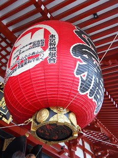
A trip to the old part of Tokyo, Asakusa!

Kaminarimon, Asakusa

Sensō-ji, Asakusa

Sensō-ji, Asakusa

Asakusa

Wednesday, August 3, 2011

To Japan's Past and Science's Future

Last week I attended an evolutionary biology conference in Kyoto.  The conference featured a wide variety of talks including:

-why our genes are encoded by DNA and not RNA
-how genes die
-the origin of mitochondria
-the stinkbug microbiome
-photosynthetic slugs
-the origin of eukaryotes
-the effect of diet and host on the primate microbiome
-horse domestication
-ancient bacterial DNA from teeth
-the effect of host living temperature on the genome
-how centromeres are lost
-the environment of the ancestor of all present life


Before and after the conference, I traveled around two of Japan's previous capitals, Nara and Kyoto.  Nara was the capital of Japan from 710 to 784.  A deer is said to have led a god to find and establish Nara as the capital and so deer are protected animals found all over Nara Park.


Nara Park

Along Nara Park, the largest wooden building in the world with the world's largest bronze statue of Buddha are found.

Todai-ji

Daibutsu


Kyoto was the capital from 1180-1868.  Kyoto is a city rich in culture, art, and food.  Many of its temples and shrines are world relics and frequently appear in film.

Mt. Daimonji

This character, dai, is one of five sites set afire on August 15th to honor the spirit world.


Fushimi Inari Shrine

Fushimi Inari Shrine is the central shrine to the god Inari in Japan.  This shrine grounds are covered in some thousand red torii gates.  I spent 2 hours walking through torii gates!


Kiyomizu-dera

Kiyomizu-dera is the temple of "clear water" situated above the trees, overlooking the city.  It is said that if one jumps and survives the 13 meter fall from Kiyomizu-dera, their wish will be granted.

I didn't jump... but I did have my wish granted of experiencing Kyoto kaiseki.

Kikunoi kaiseki first course

Kaiseki is traditional Japanese court cuisine similar to the western tasting menu.  The meal runs about 8 to 14 courses with each course featuring seasonal and exquisite foods presented in elegance and modesty.  A kaiseki experience leaves you filling reborn and heavenly.

Wednesday, July 20, 2011

How to make more DNA

After isolating a single cell, the next step is some kind of DNA analysis.  That means you need DNA.  Yes a single cell has DNA but the amount of DNA in a single cell isn't enough for most types of analysis.  So, you need to make more of that cell's DNA. 

The way to make more DNA from a single cell is to perform whole genome amplification.  Each time a cell divides, it performs essentially whole genome amplification to ensure that the newly made cell has its own genome.  This whole genome amplification takes place within the cell using its own suite of enzymes.  We have to perform a whole genome amplification in a tube where the entire contents of the cell have been spilled out. 

Currently, the best way to do whole genome amplification in a tube is by using the enzyme Phi29 DNA polymerase.  A DNA polymerase is an enzyme that makes more DNA.  Phi29 is a specific DNA polymerase that replicates by a process called strand displacement.   

http://en.wikipedia.org/wiki/File:MDA_reaction_1.JPG 


In strand displacement, random primers tell the polymerase where to begin making DNA.  The polymerase amplifies DNA for about 7 to 10 kb or until reaching a previously amplified region.  When the polymerase hits this previously amplified region, it actually displaces it and continues to make more DNA.  Meanwhile, new primers and new polymerases can begin amplifying DNA using either the original DNA or the newly replicated DNA.  The result is the branching network of DNA molecules shown in the picture above. 

The advantage of strand displacement is that it does a better job of replicating the entire genome uniformly and to high copy number than other amplification methods. 

At the very end of the entire process, all of the branching molecules are cut so that only linear molecules remain.  These molecules are then ready for DNA analysis!


FOOD!


Kinmedai (golden eye snapper)
Squid ink noodles

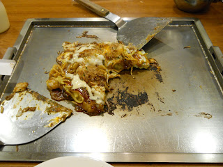
Modanyaki (savory pancake with noodles)
Sashimi
Mont blanc (chestnut cream dessert)

Wednesday, July 13, 2011

There's more than one way to dissect out a microbe.

Micromanipulation is just one way to isolate a single microbe.
This past week, I learned about two other methods used for single cell genomics.


The first was laser microdissection.


Laser microdissection is exactly what it sounds like.  You use a laser to cut out the cell you want.

http://www.leica-microsystems.com/products/light-microscopes/life-science-research/laser-microdissection/details/product/leica-lmd7000/

The part you cut out literally falls down onto a collection plate.  This might sound hard but it's not.  The laser is extremely precise and very easy to control using its computer software.  What the microscope sees is shown on the computer screen.

Find your cell of interest.

Then you just use your mouse to draw around what you want to cut out, tell the laser to go, and it's done!

Draw a circle around it.

Tell the laser to cut it out.

The collection plate with the part cut out.


While it took me about 15 minutes to micromanipulate a single cell, it took me only a few minutes with the laser dissector. 


The second technique for single cell genomics I learned about was cell sorting.


The cell sorter is a device that forces cells to move one at a time past a laser beam.  The device then records how long the cell took move past the laser and how the light from laser was refracted by the cell.  These pieces of information can be related back to the size, shape, and quality of the cell.  Most of the time, the cells being sorted have been made to fluoresce.  So the device will also measure the fluorescence coming off of the cells themselves.  Together, a fingerprint for each cell type is created.  So from a mixed population of cells, the device can type each cell and sort them apart.


Before sorting

After sorting


Cell sorting is an extremely powerful yet difficult method.  My lab was able to sort 200,000 cells in a matter of minutes.  However, it took us 8 hours to set up the device to read and sort the cells the way we wanted.  Additionally, we must do further checks to confirm the cells were correctly sorted.


The ocean, mountains, and a castle!


Pacific Ocean on the left, Mt. Fuji straight ahead

Odawara Castle

Odawara Castle

Odawara Castle